Gels formed from different gelation methods can be observed via confocal microscopy. These gels can be formed in most vessels that have a glass bottom, such as on a standard microscope slide or in a custom vessel adhered to a glass slide. Usually the easiest way is to form a gel in the circular well plate of a small cell culture dish. We typically use Greiner CELLview dishes.
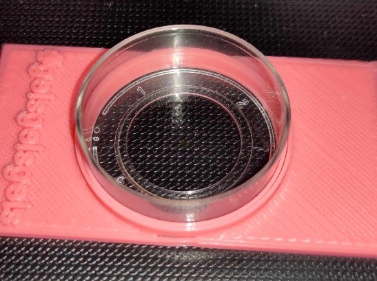
These hold ~400-500 μL of sample in their well, so gels are made at 400 μL volumes accordingly. Dishes must be thoroughly cleaned beforehand and care taken when making solvent triggered gels to limit the exposure of incompatible organic solvents to the plastic walls and lids of the dishes.
To prepare:
- Determine a suitable fluorescent dye for imaging. We typically use Nile Blue A, but other dyes, such as Nile Red or Thioflavin T, can be used. It is important to consider the hydrophobicity of the dye, as well as emission wavelengths to ensure compatibility with the gel network to be imaged and the limitations of the microscope available. Nile Blue is usually added to the aqueous portion of the gelator solutions, but others may necessitate addition to the organic solvent instead, if using solvent triggered gels. Typically we add 2 μL of a 0.1 wt% aqueous dye solution per mL of gel.
- Form gel to be imaged in confocal dish. For pH or salt/media triggered gels, this typically involves adding the trigger then quickly pipetting/ pouring the gel into centre of the dishes well. For solvent triggered gels, the organic solvent gelator solution is usually pipetted in first, spread to ensure even coverage and then followed by the addition of the water aliquot via pipette. Water should be added at an appropriate speed at the centre of the well to promote sample homogeneity. Below is a link to a video showing this step.
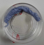
- Samples should be left to gel for an appropriate amount of time. If overnight gelation is needed, a small piece of rolled blue roll saturated with water can be carefully placed in the outer divot of the dish with tweezers to avoid the sample drying out. The dishes should then have lids fitted and be sealed carefully with parafilm to avoid evaporation of solvent.
- Samples should remain sealed until just before imaging is to be carried out.
- Imaging should be carried out away from the centre of the formed gel, to avoid any potential disturbances from the pipetting during gelation.
